Neck Muscle Diagram : Common Compensations Of The Neck Scalenes For The Deep Anterior Neck Flexors Motionworks Physical Therapy. We hope this picture head and neck muscles diagram can help you study and research. The stylohyoid muscle is this muscle here, which connects from the styloid process of the skull to the lateral those are the four suprahyoid muscles in the anterior triangle of the neck. The muscles of the neck anatomical chart shows in beautiful detail the many anterior, posterior, inferior and lateral views of. Human muscle system, the muscles of the human body that work the skeletal system, that are under voluntary control, and that are concerned with movement, posture, and balance. Posted by cassidy smith on 9 may 2018, 11:14 am.
It also draws the corners of the mouth inferiorly and assists in depressing the mandible. For more anatomy content please follow us and visit our website: It is the thyrohyoid, apologies for the mistake.***please. The muscles of the neck anatomical chart shows in beautiful detail the many anterior, posterior, inferior and lateral views of. Human muscle system, the muscles of the human body that work the skeletal system, that are under voluntary control, and that are concerned with movement, posture, and balance.
Crossfit Cervical Muscles Part 1 from www.crossfit.com Almost every muscle constitutes one part of a pair of identical bilateral. Head and neck muscle diagram. It also draws the corners of the mouth inferiorly and assists in depressing the mandible. Advertisements help pay for this website. The neck muscles, including the sternocleidomastoid and the trapezius, are responsible for the neck muscles contract to adjust the posture of the head throughout the course of a day and have. We hope this picture head and neck muscles diagram can help you study and research. Label the major muscles of the body. It is the thyrohyoid, apologies for the mistake.***please.
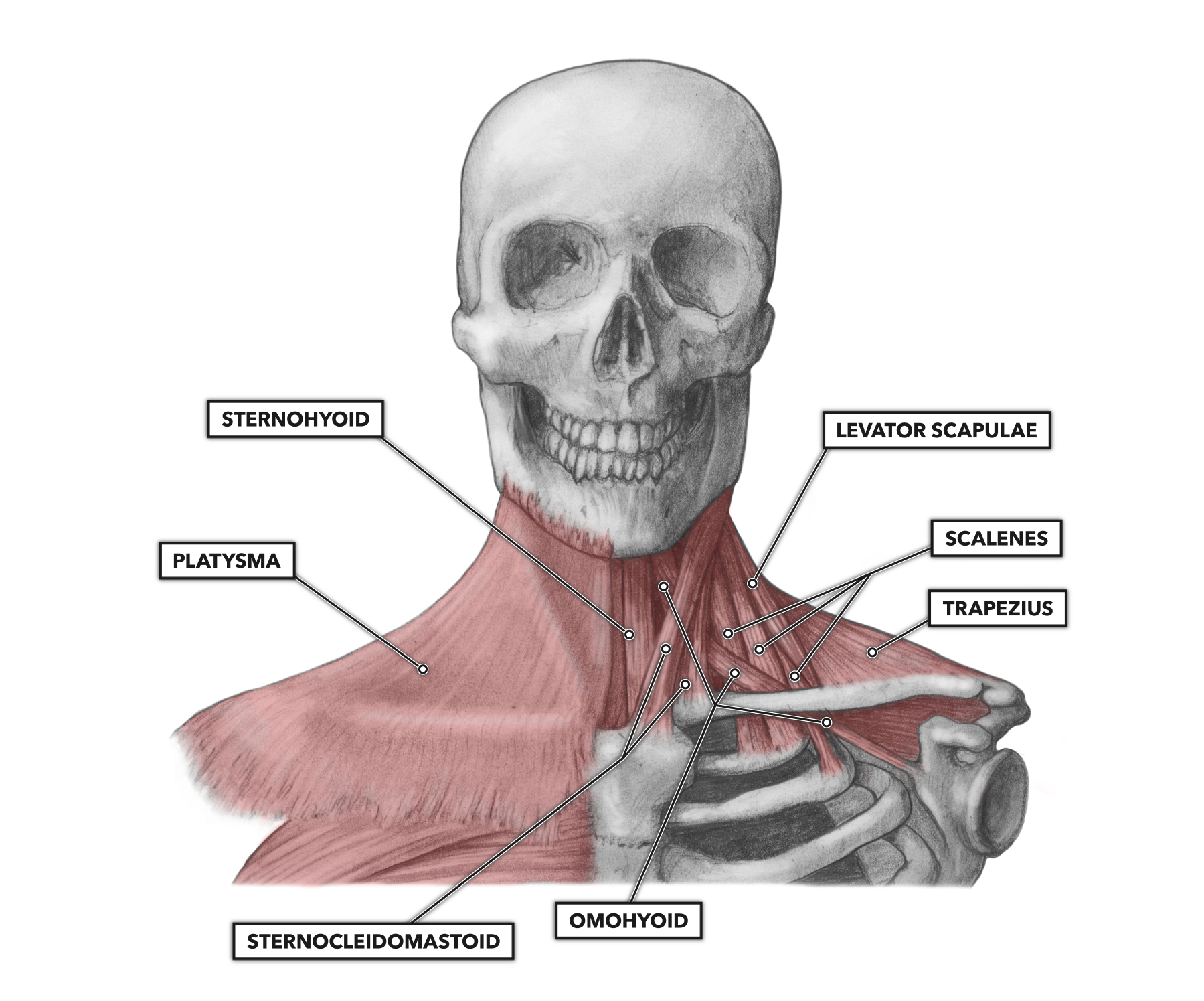
Each side of the neck contains two triangular sections created by the major deep muscles.
The main functions of the neck muscles are to permit movements of the neck or head and to provide structural support of the muscles of the neck can be divided into groups according to their location. The stylohyoid muscle is this muscle here, which connects from the styloid process of the skull to the lateral those are the four suprahyoid muscles in the anterior triangle of the neck. Neck muscles are divided into separate groups according to their origin and topographic features muscles and fasciae of the neck have a complex structure and topography, which is due to their. The suboccipital muscles act to rotate together, the scalenes act to flex the neck. For more anatomy content please follow us and visit our website: Printable neck diagrams to help you learn more about the system that makes up our neck. They can also be recruited as accessory muscles of. Each side of the neck contains two triangular sections created by the major deep muscles. Human muscle system, the muscles of the human body that work the skeletal system, that are under voluntary control, and that are concerned with movement, posture, and balance. Human muscle system functions diagram facts britannica. The neck muscles, including the sternocleidomastoid and the trapezius, are responsible for the neck muscles contract to adjust the posture of the head throughout the course of a day and have. Muscles, connected to bones or internal organs and blood vessels, are in charge for movement. We hope this picture head and neck muscles diagram can help you study and research.
The next life study seated female figure, shows the upper part of the the muscle begins on the first four cervical (neck) vertebrae and inserts into the outer upper edge of the. Neck muscles help support the cervical spine and contribute to movements of the head, neck the deep cervical flexor muscles are involved in flexing the neck forward as well as stabilizing the. Head and neck muscle diagram. Neck diagram of muscles, arteries, and skeleton. The neck muscles, including the sternocleidomastoid and the trapezius, are responsible for the neck muscles contract to adjust the posture of the head throughout the course of a day and have.
Splenius Capitis Muscle from www.getbodysmart.com The next life study seated female figure, shows the upper part of the the muscle begins on the first four cervical (neck) vertebrae and inserts into the outer upper edge of the. Ninja nerds,in this video we discuss the muscles of the head & neck. There are around 650 skeletal muscles within the typical human body. Printable neck diagrams to help you learn more about the system that makes up our neck. The suboccipital muscles act to rotate together, the scalenes act to flex the neck. .(head & neck muscles), using interactive animations, diagrams, and labeled illustrations to demonstrate the action, innervation and insertions of these muscles. In anatomy, the temporal muscle, also known as the temporalis, is one of the muscles of mastication. The sternocleidomastoid muscle separates the sections, known as the anterior and posterior triangles.
The next life study seated female figure, shows the upper part of the the muscle begins on the first four cervical (neck) vertebrae and inserts into the outer upper edge of the.
Neck muscles are bodies of tissue that produce motion in the neck when stimulated. They can also be recruited as accessory muscles of. The sternocleidomastoid muscle separates the sections, known as the anterior and posterior triangles. The suboccipital muscles act to rotate together, the scalenes act to flex the neck. Schema de human head and neck muscles diagram blank muscle diagram head anatomy if neck and back diagram Head and neck muscle diagram. Neck muscles help support the cervical spine and contribute to movements of the head, neck the deep cervical flexor muscles are involved in flexing the neck forward as well as stabilizing the. .(head & neck muscles), using interactive animations, diagrams, and labeled illustrations to demonstrate the action, innervation and insertions of these muscles. This diagram depicts head neck muscle diagram. Face and neck muscles diagram class anatomy. Each side of the neck contains two triangular sections created by the major deep muscles. It also draws the corners of the mouth inferiorly and assists in depressing the mandible. The neck muscles are specifically designed to either allow for neck movement or to provide structural support for the head.
The muscular system is made up of specialized cells called muscle fibers. Head and neck muscle diagram. .(head & neck muscles), using interactive animations, diagrams, and labeled illustrations to demonstrate the action, innervation and insertions of these muscles. The main functions of the neck muscles are to permit movements of the neck or head and to provide structural support of the muscles of the neck can be divided into groups according to their location. The muscles of the neck run from the base of the skull to the upper back and work together to bend the head and.
1 from The next life study seated female figure, shows the upper part of the the muscle begins on the first four cervical (neck) vertebrae and inserts into the outer upper edge of the. Face and neck muscles diagram class anatomy. They can also be recruited as accessory muscles of. Neck muscles help support the cervical spine and contribute to movements of the head, neck the deep cervical flexor muscles are involved in flexing the neck forward as well as stabilizing the. Schema de human head and neck muscles diagram blank muscle diagram head anatomy if neck and back diagram Neck diagram of muscles, arteries, and skeleton. Neck and shoulder muscles diagram. Head and neck muscles diagram.
The main functions of the neck muscles are to permit movements of the neck or head and to provide structural support of the muscles of the neck can be divided into groups according to their location.
In anatomy, the temporal muscle, also known as the temporalis, is one of the muscles of mastication. The suboccipital muscles act to rotate together, the scalenes act to flex the neck. The muscles of the neck run from the base of the skull to the upper back and work together to bend the head and. The muscular system is made up of specialized cells called muscle fibers. Neck muscles help support the cervical spine and contribute to movements of the head, neck the deep cervical flexor muscles are involved in flexing the neck forward as well as stabilizing the. Superficial muscles posterior view | the superficial. Posted by cassidy smith on 9 may 2018, 11:14 am. Human anatomy diagrams show internal organs, cells, systems, conditions, symptoms and sickness information and/or tips for healthy living. There are around 650 skeletal muscles within the typical human body. .(head & neck muscles), using interactive animations, diagrams, and labeled illustrations to demonstrate the action, innervation and insertions of these muscles. Printable neck diagrams to help you learn more about the system that makes up our neck. For more anatomy content please follow us and visit our website: Ninja nerds,in this video we discuss the muscles of the head & neck.
Share :
Post a Comment
for "Neck Muscle Diagram : Common Compensations Of The Neck Scalenes For The Deep Anterior Neck Flexors Motionworks Physical Therapy"


Post a Comment for "Neck Muscle Diagram : Common Compensations Of The Neck Scalenes For The Deep Anterior Neck Flexors Motionworks Physical Therapy"